Fungal Disease-
-Candidiasis-

-Candidia infections can vary from minor localized mucocutaneous infectious to widespread candidia dissemination
-Candidia is considered normal flora in the gastrointestinal and genitourinary tract, candidia can cause disease when there is an imbalance in the body's ecosystem
-Candidia can cause numerous infections: oral candidia, esophagitis, vulvovaginitis, balanitis, chronic mucocutaneous candidia, mastitis, candidemia, hepatosplenic or chronic disseminated candidiasis, urinary tract infection, endophthalmmitis, osteoarticular infections, meningitis, endocarditis, peritonitis, pneumonia, mediastinitis, pericarditis, and gastrointestinal tract colonization.
-Oral candidia is common in young infants and older adults with dentures, treated with antibiotics, chemotherapy, radiation therapy to head and neck, inhaled glucocorticoids, or patients with cellular immune problems such as HIV.
-Oral candidia presents with white plaques on the buccal mucosa, palate, tongue or oropharynx.
-Oral candidia is treated with nystatin oral suspension
-Candidia Esophagitis is most common in HIV infected patients. It is considered an AIDS defining illness. Candidia Esophagitis can also be seen in hematologic malignancies.
-Symptoms of candidia esophagitis include: odynophagia and localize their pain to the retrosternal area. Diagnosis is made by endoscopy
-Candidia vulvovaginitis is the most common form of candida. It occurs with high estrogen levels, oral contraception use, pregnancy, antibiotics, glucocorticoids, diabetes, HIV infection, IUD, and diaphragm use are risk factors
-Candidia vulvovaginitis can be diagnosed clinically but confirmed with wet mount or KOH prep. Can be treated with antifungal vaginal suppositories or diflucan
-Balanitis presents with white patches on the penis associated with burning and itching
-Mastitis-lactating women with injured nipples are at risk for developing cellulitis and also can get fungal infections also
-Invasive focal or system infection are associated with candidemia that occurs with immunosuppressed patients
-Fungal UTI usually come with hospitalized patients Can also get from a disseminated infection from the kidney
-Endophthalmitis can develop from direct eye trauma or eye surgery. Can get hematogenous spread to the retina but unusual
-Candidia Osteoarticular infections come from result from hematogenous spread or inoculation during trauma or injections
-Candidia Meningitis and endocarditis can present from hematogenous spread. Both are unusual
-Peritonitis fungal infections can occur following GI tract perforation, anastomosis leaks after bowel surgery, and necrotizing pancreatitis
-Candidia empyema occurs commonly in patients with malignancy
-Candidia mediastinitis occurs after thoracic surgery procedures. Uncommon
-Cryptococcosis-

-Cryptococcus is a fungal infection caused by cryptococcus gattii
-Cryptococcus manifests itself usually as meningoencephalitis and/or pulmonary infection
-Systemic features include fever, chills, and weight loss. Other signs and symptoms include papilledema and will have a cough with pulmonary infections
-Cryptococcus may lead to increased ICP, severe headache, hydrocephalus, ataxia, vision and hearing loss
-Workup should include CT scan of chest (for pulmonary) and head for neurologic. Also need CSF examination, funduscopic exam, and serum cryptococcal antigen
-Most patients with cryptococcal meningitis are immunocompromised. Forms of immunosuppression seen in include HIV, glucocorticoid therapy, solid organ transplantation, cancer, and hepatic failure
-Treatment for cryptococcus include antifungal agents such as amphotericin B, oral flucytosine, or oral fluconazole
-Persons with AIDS related cryptococcal meningitis need to be on maintenance therapy to prevent relapse
-Histoplasmosis-

-Histoplasmosis is a common fungal infection that is usually asymptomatic but occasionally will result in severe illness
-Histoplasmosis is usually endemic to North and Central America
-The most common areas affected by histoplasmosis is Ohio and Mississippi River valleys.
-Histoplasmosis should be considered during any of the following clinical situations: pneumonia with hilar and mediastinal lymphadenopathy, mediastinal or hilar masses, pulmonary nodules, cavitary lung disease, pericarditis with mediastinal lymphadenopathy, pulmonary manifestations with arthritis plus erythema nodosum, dysphagia with esophageal narrowing, and superior vena cava syndrome or obstruction of other mediastinal structures
-Stains for fungi, cultures, antigen detection, and serologic tests for Histoplasmosis specific antibodies can help make the diagnosis
-Can also make the diagnosis from biopsy of pulmonary tissue
-Cultures are most useful in patients with chronic pulmonary histoplasmosis
-Patients with acute diffuse pulmonary disease serologic antigen provides the highest sensitivity
-Patients with localized pulmonary disease have a lower yield on serologic or urine antigen and pulmonary cultures maybe higher yield
-Acute pulmonary histoplasmosis moderate to severe should be treated with Amphotericin B IV then followed by Itraconazole. Solumedrol should also be given
-Acute pulmonary mild to moderate histoplasmosis with symptoms greater than 4 weeks, chronic cavitary pulmonary histoplasmosis, and symptomatic mediastinal granuloma should be treated with Itraconazole
-Histoplasmosis can cause extra pulmonary syndromes including pericarditis and rheumatology syndromes
-Pneumocystis-
-Pneumocystis Jirovecii (Pneumocystis Carinii) is the organism that leads to pneumocystis carinii pneumonia (PCP)
-The incidence of PCP has decreased because of effective antiviral therapy with HIV
-PCP is one of the leading causes of opportunistic infections in the HIV infected patients with low CD4 counts or those not HIV infected patients unaware of the infection or those non compliant with therapy
-The transmission of pneumocystis is the airborne route. Primary infection occurs early in life, with 75 percent of humans infected by 4 years of age. It was believed that pneumocystis remained latent unless immunosuppressed.
-Phagocytosis, respiratory burst, and inflammatory activation of alveolar macrophages are impaired in HIV infected patients and contribute to the pathogenies of the infection
-PCP presents with fever, cough, and dyspnea presenting over days to weeks
-As CD4 counts decreases the infection rates of PCP increases
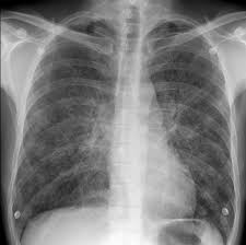
-Chest X Rays can be normal in 25 percent of patients with PCP. If negative consider CT scan of chest.
-If patient cannot have a CT scan of the chest, Gallium 67 Citrate scanning is sometimes used to screen for PCP
-Definitive diagnosis of PCP requires visualization of the cystic or trophic forms in respiratory secretions
-Pneumocystis cannot be cultured
-Immunofluorescent staining with fluorescein labeled monoclonal antibodies is the gold standard for PCP
-It is recommended to treat PCP with bactrim in non HIV infected patients
-Alternative therapies include pentamidine, atovaquone, bactrim plus dapsone, and primaquine plus clindamycin
-Patients should be treated for 21 days
-Candidia vulvovaginitis can be diagnosed clinically but confirmed with wet mount or KOH prep. Can be treated with antifungal vaginal suppositories or diflucan
-Balanitis presents with white patches on the penis associated with burning and itching
-Mastitis-lactating women with injured nipples are at risk for developing cellulitis and also can get fungal infections also
-Invasive focal or system infection are associated with candidemia that occurs with immunosuppressed patients
-Fungal UTI usually come with hospitalized patients Can also get from a disseminated infection from the kidney
-Endophthalmitis can develop from direct eye trauma or eye surgery. Can get hematogenous spread to the retina but unusual
-Candidia Osteoarticular infections come from result from hematogenous spread or inoculation during trauma or injections
-Candidia Meningitis and endocarditis can present from hematogenous spread. Both are unusual
-Peritonitis fungal infections can occur following GI tract perforation, anastomosis leaks after bowel surgery, and necrotizing pancreatitis
-Candidia empyema occurs commonly in patients with malignancy
-Candidia mediastinitis occurs after thoracic surgery procedures. Uncommon
-Cryptococcosis-
-Cryptococcus is a fungal infection caused by cryptococcus gattii
-Cryptococcus manifests itself usually as meningoencephalitis and/or pulmonary infection
-Systemic features include fever, chills, and weight loss. Other signs and symptoms include papilledema and will have a cough with pulmonary infections
-Cryptococcus may lead to increased ICP, severe headache, hydrocephalus, ataxia, vision and hearing loss
-Workup should include CT scan of chest (for pulmonary) and head for neurologic. Also need CSF examination, funduscopic exam, and serum cryptococcal antigen
-Most patients with cryptococcal meningitis are immunocompromised. Forms of immunosuppression seen in include HIV, glucocorticoid therapy, solid organ transplantation, cancer, and hepatic failure
-Treatment for cryptococcus include antifungal agents such as amphotericin B, oral flucytosine, or oral fluconazole
-Persons with AIDS related cryptococcal meningitis need to be on maintenance therapy to prevent relapse
-Histoplasmosis-
-Histoplasmosis is a common fungal infection that is usually asymptomatic but occasionally will result in severe illness
-Histoplasmosis is usually endemic to North and Central America
-The most common areas affected by histoplasmosis is Ohio and Mississippi River valleys.
-Histoplasmosis should be considered during any of the following clinical situations: pneumonia with hilar and mediastinal lymphadenopathy, mediastinal or hilar masses, pulmonary nodules, cavitary lung disease, pericarditis with mediastinal lymphadenopathy, pulmonary manifestations with arthritis plus erythema nodosum, dysphagia with esophageal narrowing, and superior vena cava syndrome or obstruction of other mediastinal structures
-Stains for fungi, cultures, antigen detection, and serologic tests for Histoplasmosis specific antibodies can help make the diagnosis
-Can also make the diagnosis from biopsy of pulmonary tissue
-Cultures are most useful in patients with chronic pulmonary histoplasmosis
-Patients with acute diffuse pulmonary disease serologic antigen provides the highest sensitivity
-Patients with localized pulmonary disease have a lower yield on serologic or urine antigen and pulmonary cultures maybe higher yield
-Acute pulmonary histoplasmosis moderate to severe should be treated with Amphotericin B IV then followed by Itraconazole. Solumedrol should also be given
-Acute pulmonary mild to moderate histoplasmosis with symptoms greater than 4 weeks, chronic cavitary pulmonary histoplasmosis, and symptomatic mediastinal granuloma should be treated with Itraconazole
-Histoplasmosis can cause extra pulmonary syndromes including pericarditis and rheumatology syndromes
-Pneumocystis-
-Pneumocystis Jirovecii (Pneumocystis Carinii) is the organism that leads to pneumocystis carinii pneumonia (PCP)
-The incidence of PCP has decreased because of effective antiviral therapy with HIV
-PCP is one of the leading causes of opportunistic infections in the HIV infected patients with low CD4 counts or those not HIV infected patients unaware of the infection or those non compliant with therapy
-The transmission of pneumocystis is the airborne route. Primary infection occurs early in life, with 75 percent of humans infected by 4 years of age. It was believed that pneumocystis remained latent unless immunosuppressed.
-Phagocytosis, respiratory burst, and inflammatory activation of alveolar macrophages are impaired in HIV infected patients and contribute to the pathogenies of the infection
-PCP presents with fever, cough, and dyspnea presenting over days to weeks
-As CD4 counts decreases the infection rates of PCP increases
-Chest X Rays can be normal in 25 percent of patients with PCP. If negative consider CT scan of chest.
-If patient cannot have a CT scan of the chest, Gallium 67 Citrate scanning is sometimes used to screen for PCP
-Definitive diagnosis of PCP requires visualization of the cystic or trophic forms in respiratory secretions
-Pneumocystis cannot be cultured
-Immunofluorescent staining with fluorescein labeled monoclonal antibodies is the gold standard for PCP
-It is recommended to treat PCP with bactrim in non HIV infected patients
-Alternative therapies include pentamidine, atovaquone, bactrim plus dapsone, and primaquine plus clindamycin
-Patients should be treated for 21 days
No comments:
Post a Comment